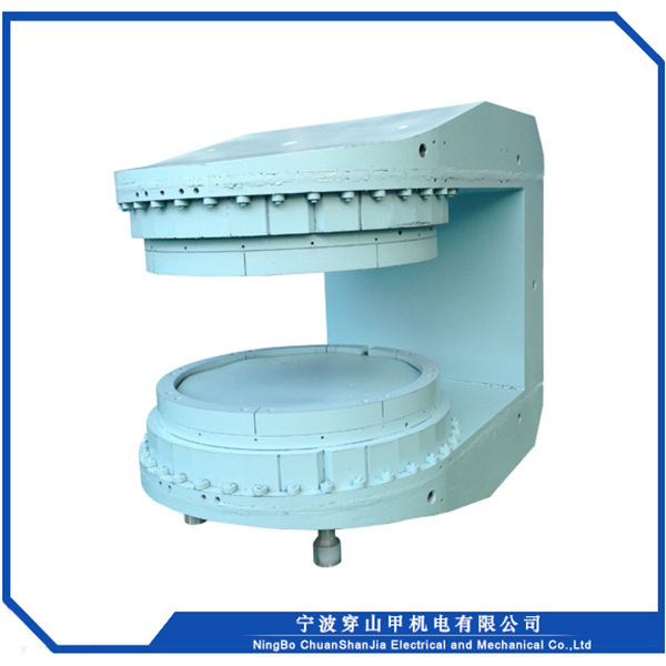
Extremity MRI
An extremity MRI is a type of scan used specifically for diagnostic imaging of the arm, leg, hand, or foot. The machine uses radio waves and a magnetic field to generate images of the inside of the extremity in order to diagnose problems with the muscles, bones, joints, nerves, or blood vessels.
Unlike a traditional MRI machine in which you need to lie still on a table for up to 60 minutes while the scanner takes a series of pictures, extremity MRI scans are much more comfortable. For this kind of MRI exam, you will simply sit in a comfortable chair and place your arm or leg in a small opening in the machine. Your head and torso will remain outside of the scanner, eliminating the claustrophobic feeling many patients experience during traditional MRI exams.
1. Use the most powerful permanent material N52 ,best open magnet design to reduce the weight.
2. Permanent magnet, no cryogens. Low maintenance costs, saving hundreds of thousands of dollars in operating costs every year
3. Open structure design, no fear of claustrophobia
4. Unique silent design, the whole scanning process is quieter and more comfortable.
5. Small part and light weight, which can meet the requirements on high-level buildings.
6. Efficient transmitting coil, SAR value is less than 1/10 of the whole body imaging system, safer and more reliable.
7. Scan in sitting, lying or weight-bearing standing positions, providing more diagnostic information.
8. Abundant 2D and 3D imaging sequences and technologies , easy using software.
9. Radio frequency coils tailored for Musculoskeletal system to improve imaging quality
10. Carefully design the positioning tool, the positioning success rate is higher, and the imaging effect is better
11.One phase AC required and low power consumption.
1.Magnetic field strength:0.3T
2.Patient gap:240mm
3.Imageable DSV: >200mm
4.Weight:<2.0Ton
5.Gradient field strength:25mT/m
6.Eddy current suppression design
7.Provide personalized customization
